Oxygen’s 8 protons attract electrons more strongly.
So “O” becomes slightly +ve and “H” becomes slightly -ve.
Causes a slight potential charge - hence the molecule is polar.
Hydrogen bonding between water molecules.
Properties of Water
Cohesion (molecules stick to each other).
Adhesion (Molecules stick to other surfaces).
Thermal Properties:
High specific heat capacity.
High latent heat of vaporisation.
High boiling point.
Universal solvent - Polar nature can interrupt intramolecular forces and cause dissociation in other molecules.
Methane vs Water
Property
Methane
Water
Formula
CH4
H2O
Molar Mass
16
18
Bonding
single covalent
single covalent
Polarity
non-polar
polar
Density (gcm-1)
0.46
1
Specific Heat Capacity (Jg-1oC-1
2.2
4.2
Latent Heat of Vaporisation (Jg-1)
760
2257
Melting Point (oC)
-182
0
Boiling Point (oC)
-160
100
Water as a Coolant
Sweat is secreted by glands in the skin.
The high specific heat of vaporisation causes the water in sweat to take away a lot of heat from the tissues in the body when evaporating.
This helps cool the body during periods of intense activity.
Similarly, dogs pant and plants transpire.
Transport in Blood Plasma
Water transports the following in blood:
Sodium Chloride - ion dissolves as Na+ and Cl-
Amino acids - polar molecules, solubility depends on R group
Glucose - polar
Oxygen - non-polar, but dissolves sparingly, haemoglobin in the blood coverts to oxyhaemoglobin to increase carrying capacity
Fat molecules - non-polar, so form lipoproteins. Lipoproteins consist of phospholipid on the outside and fat on the inside, thus helps transport.
Cholesterol - Lipoproteins, as they are non-polar
Sources
Class notes
Biology - Course Companion - Andrew Allott and David Mindorff - Oxford 2014
Enzymes
Active Sites and Enzymes
Enzymes are globular proteins that speed up reactions
They convert substrates to products
All substrates bind to the active site of its enzymes, the shape of the active site and the substrate are complementary
The shape of the active site is determined by the arrangement and bonding between amino acids that make up the enzyme
Enzyme Activity
Enzyme activity is determined by the rate of collisions between the active site and the substrate
Factors affecting enzyme activity
1) Temperature
As thermal energy increases, the particles are given more kinetic energy
So they move around more and faster, so the chances of collision increase, and enzyme activity increases
But if the temperature gets too high, the bonds between the amino acids break, altering the shape of the enzyme
So the substrate is no longer complementary to the active site and the reaction can’t be catalysed.
This is reversible upto a certain temperature, but causes permanent denaturation after that
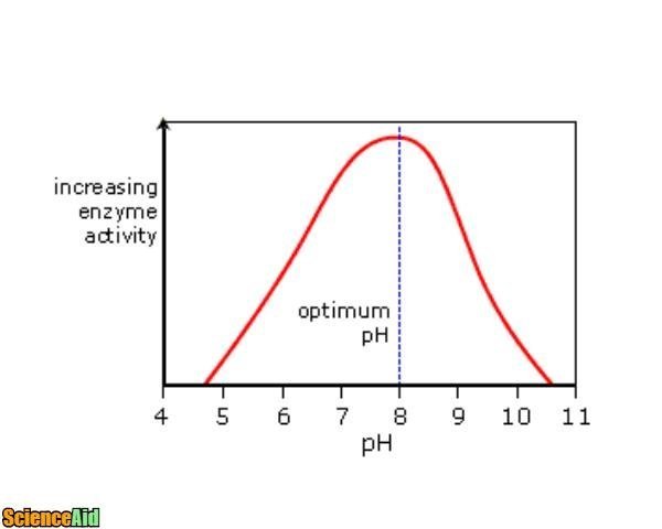
2) pH
pH is a measure of hydrogen ion concentration
Too many or too little ions can denature the enzyme by altering it’s bonding
different enzymes have different optimum pHs depending on their environment
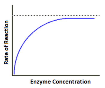
3) Substrate Concentration
If there is more substrate present, more reactions can be catalysed simultaneously, so rate of reaction would increase
But if the substrate concentration is too high, all the active sites would have already been occupied, so the rate of reaction would no longer increase
Immobilized Enzymes
Present in industries and nature
For example, on a glass surface, alginate beads, or on the wall of the villi
Advantages in industry:
Enzyme can easily be seperated from products, prevents contamination
Enzymes can be recycled easily (cost ↓)
Immobilization lowers sensitivity of enzymes to pH and temperature, so the rate at which they are degraded goes down, so can be replaced less often
Substrates are exposed to higher enzyme concentrations, so rate increases
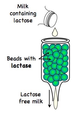
Use of Immobilized Enzymes in Producing Lactose Free Milk
Lactose is a sugar naturally present in milk
It can be broken down by the enzyme lactase
Lactose→Glucose+Galactose
Some people are lactose intolerant so cannot consume lactose
So this reaction is used to produce Lactose-Free Milk
Galactose and Glucose are sweeter than lactose, so less sugar needs to be added to dairy products made from Lactose Free Milk
Galactose and Glucose crystallize less, giving a smoother texture for ice creams
Bacteria ferment Glucose and Galactose quicker, so less time required for making yogurt
References
Class Notes
Oxford University Press BIOLOGY 2014 Edition
ScienceAid
Pathwayz
Philpot Education
DNA Replication, Transcription and Translation
CENTRAL DOGMA
Genetic Code
Universal to all organisms
Nuclear DNA consists of single copy genes + regions of highly repetitive sequences
Exons (coding sequences) and Introns (Non-Coding)
4 bases: A, T, G and C
Replication
Replication of DNA is semi-conservative, dependent on complementary base pairing
Two strands of double helix separate
Each original strand is template for new strand
Complementary nucleotides are added one by one
Since part of original strand remains, replication is semi-conservative
Complementary bases from hydrogen bonds with each other, stabilising the structure
Complementary Base Pairing: one base always pairs with another
A with T & G with C
Nature of Science: evidence for theory of semi-conservative replication
Meselson and Stahl
Used 15N, a rare isotope of nitrogen
Has 1 more neutron than 14N isotope, making it denser
Developed caesium chloride density gradient centrifugation
Method to separate the strands based on density, substance became concentrated at level corresponding to its density, creating a gradient
Cultured E. Coli, 14 generations, with 15N
Under ultraviolet light, after centrifugation, dark bands were seen
First Generation band was exactly in between original E. Coli and E. Coli with only 15N in their DNA.
Helicase
Unwinds the double helix
Separates two strands by breaking the hydrogen bonds (between complementary bases)
Uses energy from ATP
Structure: 6 globular proteins, in donut shape
Unwinding whilst separating
DNA Polymerase
Uses pre-existing strand as template
Links nucleotides together to form new strand (one at a time)
Enzyme brings nucleoside triphosphate, attaching them in the 5’ to 3’ direction
Covalent bond between phosphate group of free nucleotide and sugar of nucleotide on template strand
High degree of fidelity
Application: Polymerase Chain Reaction (PCR)
Used to make many copies of DNA sequence
If DNA heated to high temperature hydrogen bonds between complementary base pairs breaks.
When cooled, these can from again; called re-annealing
Steps (Cycle, repeated)
heated to 95 degrees celsius for 15 seconds
DNA cooled quickly to 54 degrees
Short strands of single stranded DNA present, called primers
Bind rapidly to target sequences; prevent re-annealing
Copying then starts from these primers
Enzyme TaqDNA Polymerase used
obtained from bacteria Thermus aquaticus found in hot springs
Will not denature in high temperatures
Adds nucleotides to template strands
Next cycle is started by heating to 95 degrees again
DNA is amplified
Transcription
Synthesis of mRNA copies from the DNA base sequence by RNA polymerase
RNA is single stranded; transcription occurs on only one strand
Enzyme involved: RNA Polymerase
Binds to promoter site
Moves along strand (5’ to 3’ direction) separating DNA into single strand and covalently bonding ribonucleoside triphosphate
No thymine in RNA, replaced with Uracil (U)
Once this is complete, RNA Polymerase separates from DNA strand and double helix reforms
Transcription stops at terminator site
DNA Strand that has the same base sequence is called Sense Strand
Anti-Sense Strand is what is copied from, complementary bases
Translation
synthesis of polypeptides on ribosomes
ribosomes are a binding site and catalyse the synthesis
Ribosome Structure
complex structure of proteins and ribosomal RNA
small and large subunit
1 binding site for mRNA (on small subunit)
3 binding sites for tRNA (on large subunit)
amino acid sequence determined by mRNA according to genetic code
what protein is translated depends on requirements of cell + function of cell
example: insulin secretory cells, make many copies of mRNA needed to synthesise insulin
tRNA (transfer RNA): involved in de-coding base sequence of mRNA in amino acid sequence
mRNA binds to small subunit; molecule of tRNA with anticodon complementary to first codon can be translated
second tRNA with anticodon complementary to second codon binds to ribosome active site
Ribosome transfers amino acids carried by 1st tRNA to the amino acid of 2nd tRNA by making a new peptide bond
2nd tRNA now has di-peptide
another tRNA binds with complementary anti-codon to next part of sequence
cycle continues until stop codon; then polypeptide is released
ribosome can be re-used
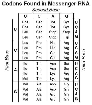
Codons and Anticodons
DNA transcribed in triplets
Codons ( 3 bases on mRNA) correspond to one amino acid in polypeptide
there are 4 bases; 20 required amino acids; therefore codon of 3 bases creates ideal possibility of 4x4x4 = 64 combinations
different codons can code for same protein
Degenerate code; reduce impact of base-substitution mutation
START CODON: AUG, promoter site
amino acid is carried on tRNA
translation depends on complementary base pairing between codons on mRNA and anticodons on tRNA
mRNA has sequence of codons that specified amino acid sequence
anticodon is complementary this codon
Cell Respiration
controlled release of energy in the form of ATP by breaking down organic compounds
one of the 7 necessary functions of life
main source is carbohydrates and lipids, but sometimes amino acids if proteins are excess in the diet.
Adenosine Tri-Phosphate
considered to be the universal energy carrier
transfers chemical energy in an immediately available form for use of metabolic processes.
releases energy by splitting ATP into ADP (di-phosphate) by hydrolysis
Structure of ATP
nucleotide derivative
purine base (Adenine) + pentose sugar (ribose) + 3 phosphates groups attached to the 5’ carbon
When energy from ATP is used in cells, its becomes converted to dissipated heat energy. Hence, continual source of ATP is required.
Anaerobic Respiration
glucose is broken down in the absence of oxygen resulting in a smaller yield of ATP.
products of anaerobic respiration vary based on organism
useful in 3 situations:
short but rapid burst of ATP/ energy
oxygen supplies have run out for the cell
environments deficient in oxygen
2.9 - Photosynthesis
Photosynthesis:- Metabolic pathway which uses carbon dioxide and water to produce carbohydrates and oxygen, in the presence of sunlight.
6CO2 + 6H2O --> C6H12O6 + 6O2
Glucose gets converted into starch and cellulose.
Leaves are green because they reflect green.
Endothermic as energy is needed to be absorbed from light (and then stored in the form of carbohydrates).
Absorption and Action Spectra
Absorption Spectrum
Shows the absorbance of light by photosynthetic pigments for all wavelengths
"Chlorophyll a**"** absorbs mostly violet and orange.
"Chlorophyll b" absorbs mostly blue and yellow.
"Carotenoids" absorb mostly blue-green and violet.
A gene is the unit of heredity. It is defined as a length of
DNA that codes for a single protein that aids in the
determination of a specific trait or characteristic of the
phenotype.
Genes occupy a specific position on one chromosome - locus
Alleles
variant form of a gene
can be two or more alleles for a gene
occupy the respective, equivalent loci on homologous chromosomes
differ from each other by slight variations in gene sequence
Homozygous - both homologous chromosomes carry identical alleles for the gene of interest Heterozygous - each homologous chromosome carries a different allele for the gene of interest
Mutation
random change in DNA base sequence
errors in copying or induced by other physical, chemical or biological agents (mutagens)
can include single base pair changes to large sections of chromosomes
gametic mutations can be inherited
somatic mutations are eliminated once the body dies.
Diseases caused by mutations Cystic fibrosis: chloride transport protein - disruption in glands that lead to symptoms like chronic lung infections Sickle Cell Anemia:
mutation in beta chain of haemoglobin (Hb) molecule
substitution of one base leading to placement of hydrophobic amino acid instead of hydrophilic one.
inherited disorder
red blood cells deform into sickle shapes, which affects movement through capillaries
reduces circulation and blocks small vessels
Genome
the whole of the genetic information in an organism
The Human Genome Project
entire base sequence of the human genes
rich mine of data
allowed for understanding of protein coding regions
“junk DNA”
comparisons with other genomes for evolutionary history
References
Class notes
Oxford University Press BIOLOGY 2014 Edition
BIOZONE Student Workbook IB Biology Second Edition
Chromosomes
Prokaryotic Chromosomes
Most prokaryotes have one naked chromosome, made out of of a single circular DNA molecule, which is present in the nucleoid. This means there is only one copy of each gene.
Plasmids:
small, extra DNA molecules
also circular and naked
consists of genes not required for basic life but for use in special circumstances (e.g. antibiotic resistance)
replicates independently of the main chromosome and can be passed between individuals through conjugation
Eukaryotic Chromosomes
linear, wound around proteins known as histones
multiple sets of chromosomes
specific positioning of the gene (loci)
homologous chromosomes - carry same sequence of genes (one is paternal and the other is maternal)
become visible during replication due to supercoiling
number of chromosomes is a characteristic feature of specie
Haploid nuclei vs. Diploid nuclei: Haploid nuclei only have one copy of each gene/ chromosome (n), while diploid nuclei have two copies of each gene/chromosome due to homologous chromosomes (2n).
Karyograms
Dividing cells are stained to produce a banding pattern on each chromosome
A micrograph is taken of the chromosomes that can be seen
Chromosomes are arranged according to size, structure, banding and position of centromere.
studied pea plants to show the inheritance of traits (specific characteristics of the individual)
proposed that inheritance was due to the transmission of discrete units -> referring to genes
Genotype: entire genetic makeup of an organism consisting of genes that influence the phenotype
Phenotype: observable characteristics of the individual as a result of the genotype
Gamete: haploid cells that are the product of meiosis and hence carry only one allele of each gene. Two gametes fuse during fertilization to form a zygote that can develop into a diploid individual.
Mendel’s Laws of Inheritance
Particulate Inheritance
Characteristics of parents are passed to offspring through discrete entities - genes
Law of Segregation
During gametic meiosis, two alleles of each gene will separate into different haploid daughter nuclei. (No gamete can have two alleles of the same gene)
Law of Independent Assortment
Allele pairs will separate independently of how other pairs are separating, in the case of unlinked genes
GENES TO BE KNOWN FOR PEA PLANT
Gene
Dominant Allele
Recessive Allele
Height of plant
Tall [TT, Tt]
Dwarf [tt]
Flower Colour
Purple [PP, Pp]
White [pp]
Flower Position
Axial [AA, Aa]
Terminal [aa]
Seed Shape
Round [RR, Rr]
Wrinkled [rr]
Seed colour
Yellow [YY, Yy]
Green [yy]
Pod Colour
Green [GG, Gg]
Yellow [gg]
Monohybrid Crosses
Cross in which the inheritance of one characteristic is observed over many generations
Dominance
Dominance: when the allele has an effect on the phenotype both in heterozygous and homozygous states Recessive: when the allele has an effect on the phenotype only in homozygous state.
Example:
Test cross: Cross that is carried out to find an unknown genotype by breeding with a homozygous recessive parent
Always remember to include genotypic and phenotypic frequencies or ratios as a conclusion for each Punnet grid/ square present.
Codominance
In codominance, neither allele is recessive to the other such that both alleles are independently expressed in the heterozygous state.
E.g.
coat colour for cattle : Red, White, Roan
coat colour for Icelandic horses: Black, White, Brown
flower colour for Mirabilis jalapa: Red, White, Pink
The intermediate phenotype is important as it is expressed in the heterozygous genotype.
Multiple Alleles
A Multiple Allele system occurs when there are more than two alleles that code for the same specific trait.
E.g. Human ABO blood group system
IA and IB are codominant.
i is recessive to both
Genotype
Phenotype
IAIA
A
IAi
A
IAIB
AB (codominance)
IBIB
B
IBi
B
ii
O (recessive)
Genetic Disorders
Present in autosomal genes (recessive) -Cystic Fibrosis Similar to regular monohybrid crosses
Present in autosomal genes (dominant) -Huntington’s Disease Similar to regular monohybrid crosses
Present in autosomal genes (codominant) -Sickle cell anaemia Intermediate phenotype is when there is a mix of regularly shaped and sickle shaped RBCs in the blood, which has been naturally selected in areas prone to malaria as the sickle cell allows for resistance against the disease, without killing the individual with sickle cell anaemia
Present in allosomal genes/ sex chromosomes -Hemophilia + Red-green colour blindness Disease allele is present on non-homologous area of the X chromosome and hence males (XY) do not have another paired allele to mask the effect of the disease allele.
Pedigree Charts
graphic illustration of inheritance patterns
Most important representations are followed universally. All pedigree charts, however, may not indicate clearly a carrier.
Mutations
radiation
mutagenic chemicals
random change to the base sequence of a gene
change the structure of the protein formed
can prevent the synthesis of protein
affects the cell in a range of intensities (e.g. cancer)
mutations in somatic (body) cells are eliminated when the individual dies
mutations in sex cells (gametes) lead to genetic disorders that can be inherited
BIOZONE Student Workbook IB Biology Second Edition
3.5 - Genetic Modification and Biotechnology
Gel Electrophoresis
Samples of DNA/protein are inserted into gel wells.
Gel is placed in conducting fluid; current is passed through.
Molecules move based off of charge.
Smaller molecules move further - can fit through gel’s pores.
Used for separation of DNA/protein fragments.
DNA is negatively charged.
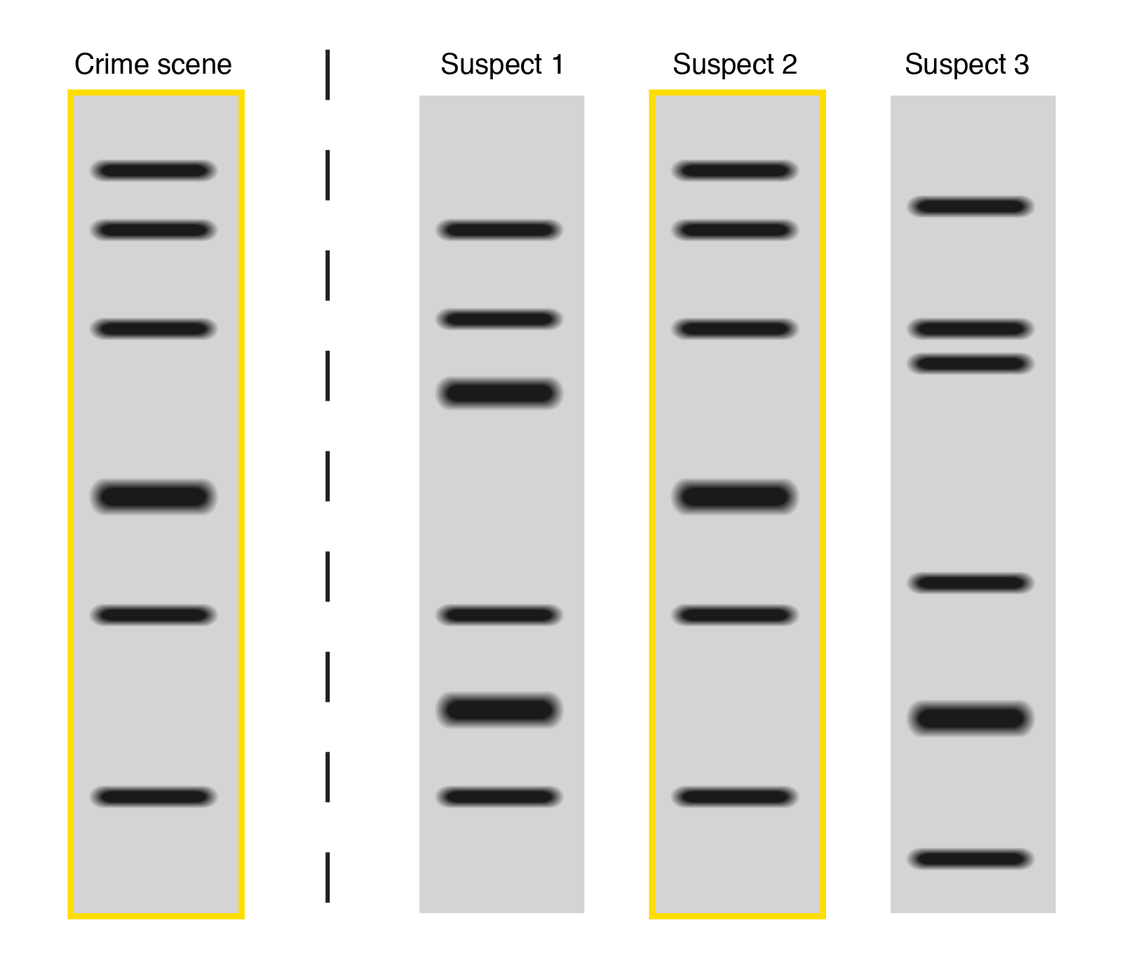
DNA Profiling
DNA samples taken and amplified through PCR
Restriction enzymes fragment DNA at specific base sequences in each sample
Fluorescent markers bind to specific base triplets in the DNA fragment so results can be seen
Samples undergo gel electrophoresis. Electric current pushes fragments along the gel
Heavier fragments stay close to origin, smaller ones move further
Banding pattern shows up for each DNA sample
Used in Forensic Investigations and Paternal Matching.
Banding Patterns shown above, source
Variable Number Tandem Repeats (VNTRs)
These are short base sequences that show variation between individuals in terms of number of repeats.
These are highly repetitive sequences - useful for DNA profiling due to uniqueness.
Genetic Modification
Genetic code is universal so genes can be transferred between species. All organisms use the same bases.
Each codon produces the same amino acid in transcription & translation, regardless of species/organism.
So the sequence of amino acids in polypeptides remains unchanged.
Gene Transfer
Restriction enzymes cut the desired gene from DNA.
Ligase joins human insulin gene to plasmid.
Recombinant plasmid is inserted back into host cell, and now expresses that gene, producing desired protein.
Transferring genes
mRNA transcript encoding desired protein obtained from eukaryotic cell. Made into cDNA (complementary DNA made with reverse transcriptase enzyme).
mRNA is easier to extract (no histones) and inserted genes are ready to be spliced - have no introns.
Bacterial enzyme and cDNA are cut with the same restriction enzymes, forming complementary sticky ends.
DNA Ligase seals eukaryotic sequence into bacterial plasmid.
Examples of Genetically Modified Organisms (GMOs)
GMO
Description
Golden Rice
Rice modified with daffodil genes to have more beta-carotene, which body converts to Vitamin A
Salt-resistant Tomatoes
Tomatoes modified to grow well in saline soils
Bt Corn
Corn modified with a bacterial insecticide gene so that it produces insect toxins within its cells
Factor IX Sheep
Sheep modified with human clotting factor IX gene so that they produce clotting factor in their milk for hemophiliacs
Round Up Ready Soy
Soybeans modified with a herbicide resistance gene so farmers can spray fields and kill weeds, not soybean plants
Rainbow Papaya
Papaya modified with viral genes that make it immune to the Papaya Ringspot Virus
Cloning
Clones; A group of genetically identical organisms.
Asexual reproduction always results in clones.
Clones are rarer in sexually reproducing organisms (eg monozygotic twins).
Clones can occur in very large numbers (eg potatoes) but can be traced to the original parent cell.
Example: A single garlic bulb will clone itself to produce many identical bulbs in growing season.
Somatic Cell Nuclear Transfer (SCNT)
Somatic cells taken from adult organism and cloned and grown in low-nutrient medium. This inactivates genes to wipe out previous patterns of differentiation
Unfertilised egg cells are taken from that species’ female and nuclei are removed
Cultured somatic cells and anucleated egg cells are placed side-by-side and zapped with a small electric pulse to fuse them together
Fused egg cells containing cell nucleus develop into embryos. Seven days later, they are implanted into a surrogate mother
Cloning low success rates, Dolly the sheep was successful 1 out of 29 times.
Sources
Biology Class notes
Biology - Course Companion - Andrew Allott and David Mindorff - Oxford 2014
Natural Selection depends on variation of phenotype, resulting in certain characteristics that are more suitable to a particular circumstance/ situation
Sources of Variation
Mutation causes new alleles of genes to be formed, enlarging gene pool
Meiosis introduces new combinations of alleles resulting in varied phenotypes, due to crossing over and independent orientation of bivalents
Sexual reproduction involves fusion of gametes from male and female. The pairing of parents introduces variation as well.
Adaptations
characteristics of an individual that make it suitable to survive in its environment
develop over time due to evolution
develop by natural selection
do not take place during one lifetime but instead over generations
Overproduction of offspring also add to the selective pressure required for natural selection and evolution to occur. There is an overall trend in living organisms to produces more number of offspring than environment can support. This leads to competition for resources and space, which makes more advantageous adaptations necessary for survival (survival of the fittest), causing these genes to be passed onto the next generation.
Better adapted individuals survive and produce more offspring than less well adapted which die/ produces less offspring.
The advantageous variation of those who survive and reproduce is inherited by offspring
Increases the frequency of these selected characteristics, decreases frequency of other alleles
Over generations, the overall characteristics of the population change to favor this selected phenotype/ allele.
Examples: Galapagos Islands: beaks of finches living in different islands AND antibiotic resistance in bacteria
local names may differ around the world, prevents international cooperation
‘science is an international venture’
consists of two parts: genus name and species name
some rules
genus: uppercase letter begins with; species name begins with lowercase letter [ Homo sapiens ]
in typed text, binomial name is in italics
after it has been used once in a text, the genus name can be abbreviated to the initial letter [ H. sapiens ]
earliest published name (from 1753 for plants or 1758 for animals) is correct
Hierarchy of Taxa
taxonomists classify species using this hierarchy
going up this hierarchy, larger numbers of species are included, and they share fewer and fewer features
use acronym: Did King Philip Come Over For Good Soup?
Feature
Bacteria
Archaea
Eukaryota
Histones associated with DNA
naked DNA, not associated with proteins
associated with proteins similar to histones
present
Presence of Introns
absent
present sometimes
present
Structure of Cell Walls
made of peptidoglycan
present, not made of peptidoglycan
sometimes present, not made of peptidoglycan
Cell Membrane
glycerol-ester lipid; side chain is unbranched; D-form of glycerol
glycerol-ester lipid; side chain is unbranched; L-form of glycerol
glycerol-ester lipid; side chain is unbranched; D-form of glycerol
archaeans are found in mostly extreme condition (far below ocean surface, hot springs etc)
viruses belong to none of these domains; not considered ‘living’ although they have genetic code
Eukaryote Classification
natural classification: genus, and accompanying taxa, consist of all the species that have evolved from one common ancestral species
convergent radiation could make organisms appear superficially related [DISADVANTAGE]
adaptive radiation could make closely related species seem different [DISADVANTAGE]
species are easier to classify (with new molecular methods, refer to 5.4) [ADVANTAGE]
features of closely related species are easy to predict as they are similar (example vestigial organs for mammals) [ADVANTAGE]
unnatural classification: species with similar features are grouped together, regardless of ancestry [birds, bats and insects classified together due to their wings]
this form of classification is misleading, as most organisms grouped together may not even be similar (apart from the feature(s) they share)
Taxonomists sometimes reclassify groups of species when new evidence shows that a previous taxon contains species that have evolved from different ancestral species.
SKILL: reading dichotomous keys
Plant Phyla
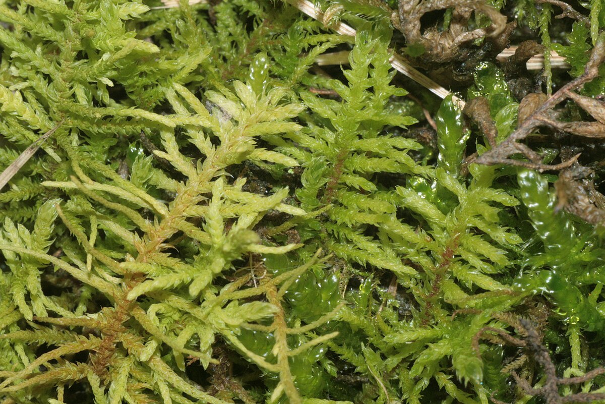
Bryophyta [relate to moss]
vegetative organs: rhizoids, but no true roots. Some have simple stem/leaves, others have thallus.
vascular tissue: no xylem or phloem
cambium: absent; no trees or shrubs
pollen not produced
no seeds produces
no ovules or ovaries
no fruits
Image
Identification
no stem, root, large leaves; usually no flowers; grow near the ground/ water sources
Filicinophyta [relate to ferns]
root, stem and leaves usually present
xylem and phloem present
cambium: absent; no trees or shrubs
pollen not produced
no seeds produces
no ovules or ovaries
no fruits
Image
Identification
leafs as fronds; never trees/shrubs
Coniferophyta [relate to conifers]
root, stem and leaves usually present
xylem and phloem present
cambium: present; allows secondary thickening of stems and development of trees and shrubs.
pollen produced in male cones
ovules in female cones
seeds produced and dispersed
no fruits
Image
Identification
presence of cone; thin sharp leaves; tree
Angiospermophyta [relate to flowering plants]
root, stem and leaves usually present
xylem and phloem present
cambium: present; allows secondary thickening of stems and development of trees and shrubs.
pollen produced by anthers
ovules enclosed in ovary of flowers
seeds produced and dispersed
fruits produces for dispersal of seeds
Image
Identification
flower, fruit, clear stem + leaves
Animal Phyla
NOTE: no diagrams necessary, only provided for understanding of anatomical differences
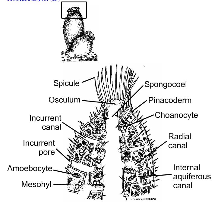
Porifera [relate to sea sponges]
no mouth or anus
no symmetry
internal skeletal needles called spicules for skeleton
pore over surface through which water is drawn for filter feeding
Image
Identification
pores over surface, irregular shape
Cnidaria [relate to jellyfish]
only mouth
radial symmetry
soft [except hard coral, CaCO3 secreted]
tentacles arranged in ring around mouth, stinging cells
Image
Identification
tentacles around mouth, no regular skeleton
Platyhelminthes [relate to flatworms]
only mouth
bi-lateral symmetry
soft, no skeleton
flat, thin bodies, shape of a ribbon; no blood system or system for gas exchange
Image
Identification
flat body, ribbon shape
Mollusca [relate to snail/octopus]
mouth and anus
bi-lateral symmetry
shell made of CaCO3
mantle, secretes the shell; rasping radula for feeding
Image
Identification
hard outer shell (exception octopus)
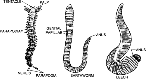
Annelida [relate to leeches]
mouth and anus
bi-lateral symmetry
internal cavity, fluid under pressure
ring-shaped segments, blood vessels often visible
Image
Identification
bristles present on body, ring like segments
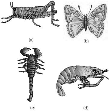
Arthropoda [relate to insects]
mouth and anus
bi-lateral symmetry
external skeleton, made of chitin
segmented body, joints between sections
Image
Identification
segmented body
Chordata (Vertebrates)
Bony ray-finned fish
Amphibians
Reptiles
Birds
Mammals
Scales which are bony plates in skin
Soft moist skin, permeable to water and gas
Impermeable skin; scales of keratin
Skin + Feathers, of keratin
Skin + Follicles with hair made of keratin
Gills covered by operculum, gill slit
Simple lungs, small folds and moist skin for gas exchange
Lungs with extensive folding (increase surface area)
Lungs with para-bronchial tubes; ventilated with air-sac
Lungs with alveoli; ventilate with diaphragm and ribs
No limbs
Tetrapods + pentadactyl limbs
Tetrapods + pentadactyl limbs
Tetrapods + pentadactyl limbs
Tetrapods + pentadactyl limbs
Fins, supported by rays
Four legs when adult
Four legs (exception: snakes)
Two legs, two wings
Four legs (or two wings/ arms and two legs)
Eggs and sperm released for external fertilisation
Eggs and sperm released for external fertilisation
Sperm passed into the female for internal fertilisation
Sperm passed into the female for internal fertilisation
Sperm passed into the female for internal fertilisation
Remain in water throughout lifecycle
Larval stage: water; adult life: usually land
Females lay eggs with soft shell
Females lay eggs with hard shell
Give birth to young live; feed with mild from mammary glands
Swim bladder containing gas for buoyancy
Eggs coated in protective jelly
Teeth of one type, no living parts
Bill/Beak no teeth
Teeth of two kinds, living core
Do not maintain constant body temperature
Do not maintain constant body temperature
Do not maintain constant body temperature
Maintain constant body temperature
Maintain constant body temperature
Cladistics
Clades
group of organisms that have evolved from a common ancestor;
species can evolve and split to form new species (speciation)
these groups share common characteristics due to common ancestor (derived characteristics)
evidence gained from base sequence of gene (or constituent amino acids of proteins)
Molecular Clocks
sequence differences accumulate gradually over time
positive correlation between number of differences between species and time since they diverged
evidence suggests the rate of mutations occurring is roughly constant, can be used to estimate time of diversion
Analogous or Homologous?
homologous structures are similar due to ancestry
pentadactyl limbs
analogous structures are similar due to convergent evolution
human eye and octopus eye (similarities in structure and function, evolved independently)
similarities in analogous and homologous structures can be misleading; for this reason morphology is rarely used to classify organisms
Cladograms
tree diagrams that show the most probable sequence of divergence in clades
when two clades branch off, it is called a node
primate cladograms
reclassification
(NOS: falsification, one theory of superseded by another)
reclassification of figwort family, using evidence of cladistics
Digestion and Absorption
system to break down large insoluble molecules, to small soluble particles that can be absorbed
requires surfactants to break up lipid droplets and enzymes to catalyse reactions
Glandular cells in the lining of the stomach and intestines produce the enzymes
Surfactants and other enzymes are secreted by accessory glands that have ducts leading to the digestive system
Controlled, selective absorption of the nutrients takes place in the small intestine and colon
Some small molecules (alcohol) diffuse through the stomach lining
(diagram necessary for drawing and labelling)
Structure
Function
Mouth
control of eating and swallowing; mechanical digestion of food by chewing and mixing saliva (lubricant); enzymes that start starch digestion
Oesophagus
movement of bolus by peristalsis from mouth to stomach
Small Intestine
final stage of digestion of lipids, carbohydrates, proteins and nucleic acids; neutralising acids from stomach; absorption of nutrients
Stomach
Churning and mixing with secreted water and acid (kills foreign bacteria and pathogens); initial stages of protein digestion
Pancreas
secretion of lipase, amylase and protease
Liver
Secretion of surfactants in bile to break up lipid droplets
Gall Bladder
Storage and regulated release of bile
Large Intestine
Re-absorption of water, further digestion of carbohydrates by symbiotic bacteria; formation and storage of faeces
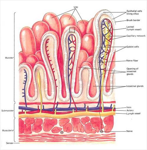
Structure of wall of Small Intestine
wall is made of layers of living tissue
four layers
serosa: outer coat
muscle layers: longitudinal muscle and (inside it) circular muscle
sub-mucosa: tissue layer with blood and lymph
mucosa: the lining of the small intestine; epithelium that absorbs nutrients on its inner surface
Peristalsis
contraction of circular and longitudinal muscle layers
in small intestine mixes the food with enzymes and helps move it
circular and longitudinal are smooth muscle; relatively short cells, not elongated fibres
exerts continuous moderate force with short periods of vigorous contraction
Contraction of circular muscles constricts the gut; prevents it from being pushed back towards the mouth (always specify direction )
Contractions are controlled unconsciously
In oesophagus, moves quickly in continuous wave
In gut, movement is slower
Peristalsis only occurs in one direction, away from the mouth
main function of peristalsis in the intestine is churning of the semi-digested food to mix it with enzymes
speed up process of digestion
Pancreatic Juice
pancreas secretes enzymes into the lumen of small intestine
pancreas contains two types of gland tissue
Small groups of cells secrete the hormones insulin and glucagon
remainder of the pancreas synthesises and secretes digestive enzymes into the gut
mediated by hormones synthesised in stomach and enteric nervous system
Small groups of gland cells cluster round tubes called ducts
digestive enzymes secreted by pancreatic gland cells on ribosomes in rER
process in Golgi Apparatus and secreted by exocytosis
ducts merge to form larger ducts; large volume secreted
Enzymes: amylase to digest starch, lipases to digest triglycerides and phospholipids and proteases to digest proteins and peptides.
Digestion in the Small Intestine
Enzymes digest most macromolecules in food into monomers in the small intestine
hydrolysis reactions
starch digested to maltose by amylase
triglycerides are digested to fatty acids and glycerol / monoglycerides by lipase
phospholipids are digested to fatty acids, glycerol and phosphate by phospholipase
proteins and polypeptides are digested to shorter peptides by protease
The wall of the small intestine produces a variety of other enzymes
most remain immobilised in the plasma membrane of epithelium cells lining
Examples include:
Substrate
Enzyme
Product(s)
DNA and RNA
nucleases
nucleotides
maltose
maltase
glucose
lactose
lactase
glucose, galactose
sucrose
sucrase
glucose, fructose
peptides
exopeptidases
dipeptide remains
dipeptides
dipeptidases
amino acids
Cellulose is not digested and passes on to the large intestine; dietary fibre
Villi and Digestion
Villi increase the surface area of epithelium over which absorption is carried out
Absorption : process of taking substances into cells and the blood is called
area is increased by the presence of villi (x10)
Villi : small finger-like projections of the mucosa
Absorption by Villi
Villi absorb monomers formed by digestion as well as mineral ions and vitamins
epithelium: permeable enough to allow useful nutrients to pass through, a barrier to harmful substances
If harmful substances pass through the epithelium they are removed from the blood and detoxified by the liver
Methods of Observation
Different methods of membrane transport are required to absorb different nutrients
The nutrients must first be absorbed into epithelium cells through the part of the plasma membrane
Examples include simple diffusion, facilitated diffusion, active transport and exocytosis
Triglycerides must be digested before they can be absorbed.
The products of digestion are fatty acids and monoglycerides, which can be absorbed into villus epithelium cells by simple diffusion ( can pass between phospholipids in the plasma membrane )
Fatty acids are also absorbed by facilitated diffusion as there are fatty acid transporters, which are proteins in the membrane
Once inside the epithelium cells, fatty acids are combined with monoglycerides to produce triglycerides, which cannot diffuse back out
Triglycerides coalesce with cholesterol to form droplets, which become coated in phospholipids and protein
These lipoprotein particles are released by exocytosis through the plasma membrane on the inner side of the villus epithelium cells.They then either enter the lacteal or enter the blood capillaries.
Glucose cannot pass through the plasma membrane by simple
diffusion because it is polar and therefore hydrophilic.
Sodium–potassium pumps in the inwards-facing part of the plasma membrane pump sodium ions by active transport from the cytoplasm to the interstitial spaces inside the villus and potassium ions in the opposite direction
Sodium–glucose co-transporter proteins in the microvilli transfer a sodium ion and a glucose molecule together from the intestinal lumen to the cytoplasm of the epithelium cells; passive but depends concentration gradient created by pumps
Glucose channels allow the glucose to move by facilitated diffusion from the cytoplasm to the interstitial spaces inside the villus and on into blood capillaries in the villus
Digestion of Starch
Starch is a macromolecule, composed of many α-glucose monomers
Must be digested in the small intestine to allow absorption
Any 1,4 bond in starch molecules can be broken by amylase
Because of the specificity of its active site, amylase cannot break 1,6 bonds in amylopectin.
Maltase, glucosidase and dextrinase digest maltose, maltotriose and dextrins into glucose
The blood in these venules is carried via the hepatic portal vein to the liver, where excess glucose can be absorbed by liver cells
NOS: Modelling Physiological Processes
dialysis tubing can be used to model absorption in the intestine
dialysis tubing made from cellulose.
pores in the tubing allow water and small molecules or ions to pass through, but not large molecules.
These properties mimic the wall of the gut: permeable to small rather than large particles.
Dialysis tubing can be used to model absorption by passive diffusion and by osmosis
It cannot model active transport and other processes that occur in living cells
Gas Exchange
Ventilation
maintains concentration gradients of oxygen and carbon dioxide between air in alveoli and blood flowing in capillaries
In humans gas exchange occurs in small air sacs called alveoli inside the lungs
Gas exchange happens by diffusion between air in the alveoli and
blood flowing in the adjacent capillaries
The gases only diffuse because there is a concentration gradient (air in the alveolus has a higher concentration of oxygen and a lower concentration of carbon dioxide than the capillary )
Process of maintaining these concentration gradients by pumping fresh air and removing stale air is called ventilation
flattened layer of thin cells (diffusion distance decreases)
dense network of capillaries
moist lining (gases can dissolve)
diffusion of oxygen down the concentration gradient
Type 1 Pneumocytes
Type I pneumocytes are extremely thin alveolar cells
Large total surface area for diffusion
The wall of each alveolus has a single layer of cells (epithelium)
Reduced diffusion distance (adaptation)
Type 2 Pneumocytes
Type II pneumocytes secrete a solution with surfactant
Creates a moist surface inside the alveoli to prevent the sides from adhering (reducing surface tension)
film of moisture allows oxygen in the alveolus to dissolve and then diffuse to the blood in the alveolar capillaries
Surfactant
structure similar to that of phospholipids
form a monolayer on the surface of the moisture
hydrophilic heads facing the water and the hydrophobic tails facing the air
reduces the surface tension
prevents the water from causing the sides of the alveoli to adhere
helps to prevent collapse of the lung
premature babies are often born with insufficient pulmonary surfactant
can suffer from infant respiratory distress syndrome
Airway for Ventilation
Air enters the ventilation system through the nose or mouth and then passes down the trachea
rings of cartilage in its wall to keep it open even when air pressure inside is low
Air is carried to the bronchi and then to the alveoli in bronchioles
bronchioles have smooth muscle fibres in their walls
Pressure Changes
pressure and volume are inversely related
Muscle contractions cause the pressure inside the thorax to drop below atmospheric pressure (air rushes into lungs, inspiration)
Muscle contractions cause the pressure inside the thorax to rise above atmospheric pressure (air rushes out lungs, expiration)
Antagonistic Muscles
Muscles do work when they contract by exerting a pulling force (tension) that causes a particular movement (become shorter)
Muscles lengthen while they are relaxing, but this happens passively (no work done)
can only cause movement in one direction
Antagonistic pair of muscles : When one muscle contracts and causes a movement, the second muscle relaxes and is elongated by the first; then the opposite happens;
Process
Muscle
Inspiration
Expiration
volume and pressure changes
N/A
Volume in thorax increases, pressure decreases
Volume in thorax decreases, pressure increases
movement of diaphragm
diaphragm
diaphragm contracts; moves downward and pushes abdomen wall out
diaphragm relaxes; can be pushes upwards into a more domes shape
abdomen wall muscles
Muscles in the abdomen wall relax allowing pressure from the diaphragm to push it out
Muscles in the abdomen wall contract pushing the abdominal organs and diaphragm upwards
movement of rib-cage
External intercostal muscles
contract, pulling the ribcage upwards and outwards
relax and are pulled back into their elongated state
Internal intercostal muscles
relax and are pulled back into their elongated state
contract, pulling the ribcage inwards and downwards
Epidemiology
Obtain evidence for theories
W.R.T understanding causes of cancer
Epidemiology is the study of the incidence and causes of disease
Based on observation, not experimentation
survey data is collected that allows the association
A correlation between a risk factor and a disease does not prove that the factor causes the disease (confounding factor)
usually necessary to collect data on many factors apart from the one being investigated
Lung Cancer
Causes of Lung Cancer
smoking
passive smoking
air pollution
radon gas
asbestos, silica
Consequences of Lung Cancer
difficulties with breathing
persistent coughing
coughing up blood
chest pain
loss of appetite/ weight loss
general fatigue
Metastasis
Surgery, Chemotherapy, Radiotherapy
Emphysema
total surface area for gas exchange is reduced
smaller number of larger air sacs with much thicker walls
lungs also become less elastic, so ventilation is more difficult
Phagocytes inside alveoli prevent lung infections by engulfing bacteria and produce elastase (protein-digesting enzyme)
An enzyme inhibitor called alpha 1-antitrypsin (A1AT) prevents elastase and other proteases from digesting lung tissue
In smokers, the number of phagocytes in the lungs increases and they produce more elastase
Genetic factors affect the quantity of A1AT produced
damage to alveoli is usually irreversible
patient lacks energy, shortness of breath
Transcription [AHL]
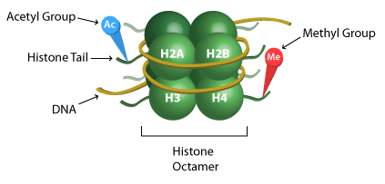
Nucleosomes + Super-Coiling
DNA in eukaryotes are associated with histone proteins
Nucleosomes Consists of octamer, DNA combination, attached to a H1 Histone
Served to protect DNA from damage and allow long lengths of DNA to be super-coiled
Supercoiling
allows chromosome to be mobile in mitosis and meiosis
supercoiled DNA cannot be transcribed for protein synthesis
allows genes to be turned on and off
DNA consists of single copy genes and regions of highly repetitive sequences (Exons, coding sequences and Introns, non-coding sequences)
While Introns were thought to be “junk DNA” they are now know to serve a purpose
production of rRNA and tRNA
genetic fingerprinting
factors that regulate gene expression
telomeres [ protective function; DNA cannot be replicated all the way to the end so telomeres prevent loss of important genes]
Regulation of Transcription
Operator: region of DNA that can regulate transcription
Promoter: non-coding DNA with function; binding site of RNA Polymerase
Gene Expression and Transcription need to be regulated to control what proteins get synthesised and what don’t
Example in Prokaryotes: lactose metabolism, negative feedback
Factors that help in these are promoters, enhancers and silencers
Both environmental factors and cellular function play a role in which proteins are synthesised
Nucleosomes in Transcription
chemical modification of the tails of histones is an important factors in determining whether a gene will be expressed
Addition of **acetyl, methyl or phosphate group **
Example: histone acetylation neutralises positive charge; allows for less condensed structures & higher levels of transcription
chemical modification of histone tails can either activate or deactivate genes by decreasing/ increasing the accessibility of the gene to transcription factors
usually addition of an acetyl group means increased transcription, while the addition of methyl group means reduced transcription
SKILL : analysing changes in DNA methylation patterns
Epigenetics
Chemical modifications are called epigenetic tags
sum of these tags are called an epigenome
provide understanding of environmental factors affecting inheritance
when 2 reproductive cells (sperm, egg cell) with epigenetic tags meet, epigenome is erased through reprogramming
1% is not erased, called imprinting
example of imprinting is gestational diabetes
Direction of Transcription
occurs in 5’ to 3’ direction
3 stages: initiation, elongation and termination
begins at promoter site; site of binding for RNA Polymerase
Operators
DNA Sequence
Binding Protein
Function
enhancer
activator
increases rate of transcription
silencer
repressor
decreases rate of transcription
promoter
RNA Polymerase
initiates transcription
Post-transcriptional Modification
eukaryotic cells modify mRNA after transcription
in prokaryotes, translation and transcription is coupled [no nuclear membrane]
cell membrane in eukaryotes allows for significant post transcriptional modification before mature transcript exits nuclear
examples of modification: removing introns
RNA splicing includes:
removing introns
splicing all/some remaining exons together
addition of 5’ cap
polyA tail added
Splicing of mRNA increases number of different proteins an organism can produce [particular exon may or not be including, increasing combinations]
Application: Morphogens
determine body patterns during embryonic development
diffuse across surface, in varied concentrations
regulate production of transcriptional factors
change rate of growth of different cells, creating ideal body shape like size of fingers, hands etc.
Translation
Component Structure
Ribosomes
Large unit (50S) and small unit (30S).
Made of proteins and ribosomal RNA (rRNA).
Three binding sites for tRNA: exit site (E), peptidyl site § and aminoacyl site (A).
Two tRNA molecules can bind at the same time.
Has a binding site for mRNA on the surface of the ribosome (for your visualisation, think lower surface).
tRNA
Have sections that become double-stranded because of complementary base-pairing.
Clover shaped with 3 loops: one is an anticodon loop.
The base sequence CCA at the 3’ end on top for attachment of amino acid.
tRNA activation
Each tRNA recognised by tRNA-activating enzymes that attaches specific amino acid to that tRNA – uses ATP.
20 different tRNAs, 20 different amino acids, and 20 different activating enzymes.
ATP and amino acids bind to enzyme.
Amino acid is activated (it gets energy) by hydrolysis of ATP (makes AMP which is adenosine monophosphate and pyrophosphate) and covalent bonding of AMP to enzyme.
tRNA binds to active site of enzyme, amino acid binds to attachment site on tRNA and AMP is released: energy released to charge tRNA.
Energy from bond later used to attach amino acid to growing peptide chain.
Initiation
assembly of components involved
small ribosomal subunit binds to mRNA at the binding site (start codon AUG)
initiator tRNA with methionine
large ribosomal subunit also binds such that initiator tRNA is on the P site
the next codon signals for the appropriate tRNA to bind to the a site.
peptide bond forms between the two amino acids
Elongation
ribosome moves down mRNA
tRNA from P site is now in the E site
tRNA from A site is now in the P site
first tRNA, now in E, is released
while a new tRNA, according to complementary base pairing joins to the A site
peptide bond forms again
series of repeated steps until the end of the mRNA
Termination
reaches a stop codon with no amino acid
polypeptide is released
Note that direction of translation is 5’ to 3’ of the mRNA strand.
Free and Bound Ribosomes
Proteins are synthesised in both ER and cytosol.
Proteins are usually needed for mitochondria or chloroplasts = free ribosomes.
Proteins needed for lysosomes, ER, Golgi apparatus, plasma membranes, outside cell = ER bound ribosomes.
Structures of Proteins
Primary structure: just peptide bonds between amino acids to form a polypeptide.
Secondary structure: alpha-helices and beta-pleated sheets due to hydrogen bonding between N-H and C-O of peptide bond.
Tertiary structure: folding of polypeptide due to interactions with R-groups: +ve and -ve charged R-groups interact, polar R-groups interact with each other, hydrophobic amino acids orient IN while hydrophilic OUT, and some R-groups form disulphide bridges.
Quaternary structure: the way polypeptides fit together; only in proteins with more than one polypeptide.
heavily dependent on oxidation and reduction reactions between compounds
substances known as electron carriers link redox by accepting and donating electrons when required
main electron carrier in respiration is nicotinamide adenine dinucleotide (NAD)
NAD++2H→NADH+H+
Phosphorylation reactions is the addition of a phosphate molecule (PO43-)
makes the organic compound less stable and more reactive i.e. activation
Hydrolysis of ATP as an exergonic reaction to release energy for couple metabolic endergonic and non-spontaneous reactions
Process
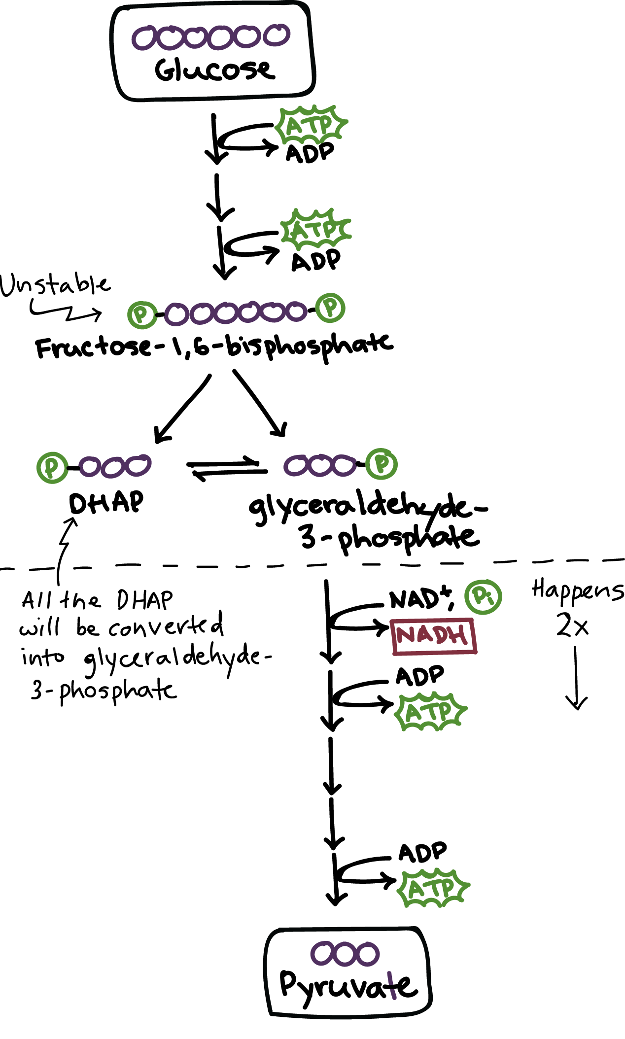
Glycolysis
occurs in the cytoplasm
does not require oxygen
has two phases: 1) energy requiring and 2) energy producing
Glucose (6C) is rearranged and phosphorylated with 2 phosphate groups to produce Fructose 1,6 biphosphate (6C). Energy is required: 2 ATP --> 2 ADP
Lysis: Fructose biphosphate is split into half: DHAP (3C) and Glycerate-3-phosphate. Glycerate-3-phosphate continues in the next step but the other compound, DHAP, is easily converted into Glycerate-3-phosphate to continue as well.
Series of reactions, oxidation by de-hydrogenation. to convert Glycerate-3-phosphate (3C) into pyruvate (3C) which releases energy. 2 ADP --> 2 ATP and NAD+ --> NADH + H+ per 3C molecule, therefore twice as much per glucose molecule
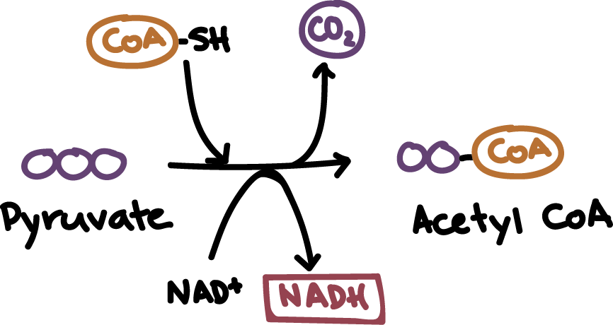
Link Reaction
occurs in the matrix of the mitochondria
does not require oxygen
links glycolysis to Kreb’s cycle
De carboxylation, therefore CO2 is released (waste), and de-hydrogenation of pyruvate molecules (3C) to form Acetyl CoA (2C). Reduces NAD+ + --> NADH + H+ per pyruvate molecule, so two molecules per glucose molecule.
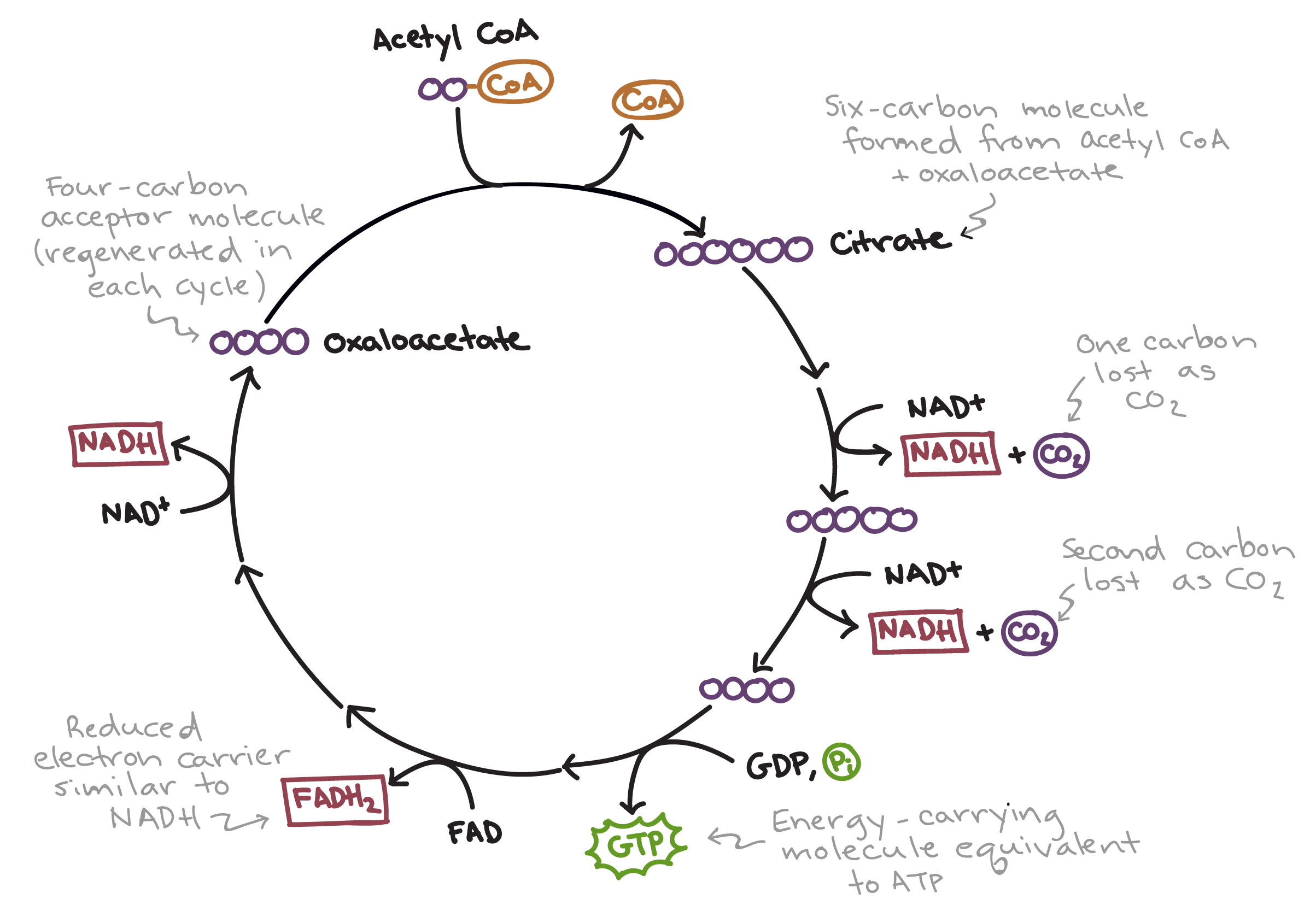
Kreb’s cycle/ Citric Acid cycle
a cyclic progression of reaction using acetyl CoA as its fuel
redox reactions release energy carried in the form of electron carriers, NADH and FADH2.
occurs in the matrix of the mitochondria
no oxygen required
runs twice per glucose molecule
Acetyla CoA (2C) combines with oxaloacetate (4C) to form citrate (6C)
Citrate (6C) is de-carboxylated into alpha ketoglutanate (5C)reducing NAD+ --> NADH + H+.
Alpha ketoglutanate (5C) is de-carboxylated into a (4C) compound, oxalosuccinate, reducing NAD+ --> NADH + H+.
This (4C) compound undergoes a series of configuration changes resulting in more release of energy and electron carriers.
NAD+ --> NADH + H+
FAD + H2 --> FADH2
ADP + Pi --> ATP
The last (4C) compound, oxaloacetate is eventually regenerated from oxalosuccinate, to continue reacting with incoming Acetyl CoA, looping the process into a cycle.
Decarboxylation produces CO2 (waste)
Electron Transport Chain
Occurs on the the cristae, folds of the inner mitochondrial membrane
transfer of electrons between proteins
coupled with proton pumping to create gradient
sets up for chemiosmosis that allows for ATP production
oxygen required
Chain of proteins arranged on the cristae in order from least electronegative to most.
Electron carrier approaches the first protein on the chain and becomes oxidised to release 2e- : NADH + H+ --> NAD+ + 2H+ + 2e-
Each electron carrier protein gets oxidized and reduced in turn, passing on electrons to the next, more electronegative, protein in the chain.
This process releases energy to pump protons, H+, from the matrix into the intermembrane space.
Due to the small volume and the membrane impermeability to protons, a concentration gradient of protons build up quickly with the high concentration in the intermembrane space.
Oxygen, being the most electronegative when compared to the protein, is present in the matrix, is the final electron acceptor.
Chemiosmosis
diffusion of the protons
catalyses oxidative phosphorylation of ADP into ATP
final stage
also requires oxygen
occurs across the inner mitochondrial membrane and in the matrix
Protons move done concentration gradient from within intermembrane space into matrix through specific channel proteins
The channel proteins, termed ATPase, also acts as an enzyme that is activated by the passage of the protons.
Head of the protein rotates, allowing Pi to bind to it, catalysing the phosphorylation: ADP + Pi --> ATP
The reduced oxygen in the matrix is needed to bind with the incoming free protons, forming water (waste product). This maintains concentration gradient.
Yield
Each NADH run by the ETC ~= 3 ATP
Each FADH2 tun by the ETC ~= 2 ATP
Glycolysis
Link
Kreb’s Cycle
ATP equivalent totally
ATP
2
x
2
2
NADH
2
2
6
30
FADH2
x
x
2
4
Total: 38 ATP per glucose molecule broken down in aerobic respiration.
Structure and Function of Mitochondria
Outer membrane : separates contents from rest of cell for a compartments with ideal conditions
consists of light dependent and light independent reactions
takes place in chloroplast in leaf cells
Light Dependent
inter-membrane space of thylakoids
produces reduced NADP and ATP
convert light energy to chemical energy
3 overall steps
excitation of light system by light (photo-activation)
production of ATP through electron transport chain
reduction of NADP+
Photo-activation
absorption of light by photosystems generates excited electrons
light harvesting arrays (in thylakoids) + reaction centres
Photosystem II is the first to be activated ( 680nm wavelength of light); Photosystem I (700 nm wavelength)
Photolysis
excited electrons from PS II are transferred to an electron transport chain;
electron acceptor in this chain is called plastoquinone
as they pass through the chain, lose energy, used to move H+ into the lumen of thylakoid from stroma
splitting of water molecule to generate electrons in LDR
chlorophyll is not powerful oxidising agent
2H2O --> O2 + 4H+ + 4e- [water-splitting enzyme]
oxygen waste product, diffuses out of leaf
When the electrons reach the end of the chain of carriers they are passed to plastocyanin, a water-soluble electron acceptor in the fluid
Production of NADPH
Photosystem I, at reaction centre, excited electrons move down their own electron transport change
energy used by NADP reductase (enzyme)
reduces NADP to NADPH, into stroma
electron for PS I comes from plastocyanin; non cyclic photo-phosphorylation
Production of ATP (Photophosphorylation)
H+ ions will diffuse passively through ATP synthase due to gradient built up
chemiosmosis (higher to lower concentration, from thylakoid space to stroma)
ATP synthase is channel + enzyme; rotating head creates energy for phosphorylation
ADP converted to ATP
Light Independent Reaction
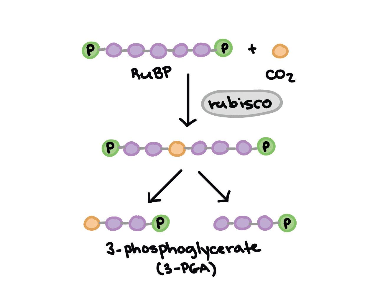
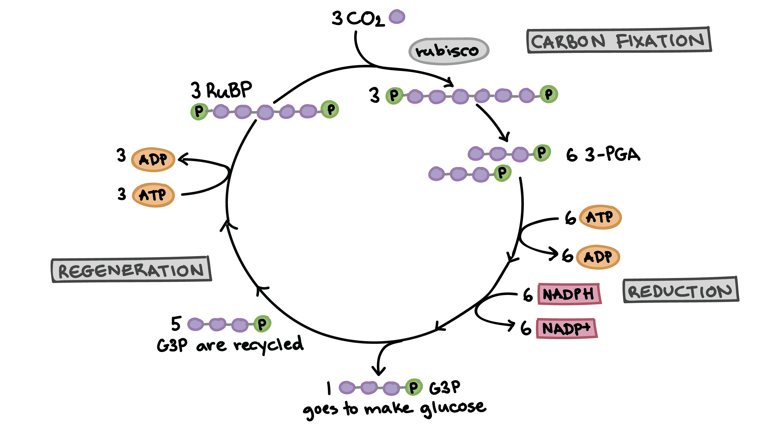
Calvin’s Cycle (Stroma)
3 RuBP (5C) (Rubisco Bisphosphate) undergo carbon fixation due to rubisco (enzyme)
This compound formed (6C) (name not necessary) is split in half to form 6 Glycerate-3- Phosphate (labelled 3-PGA)
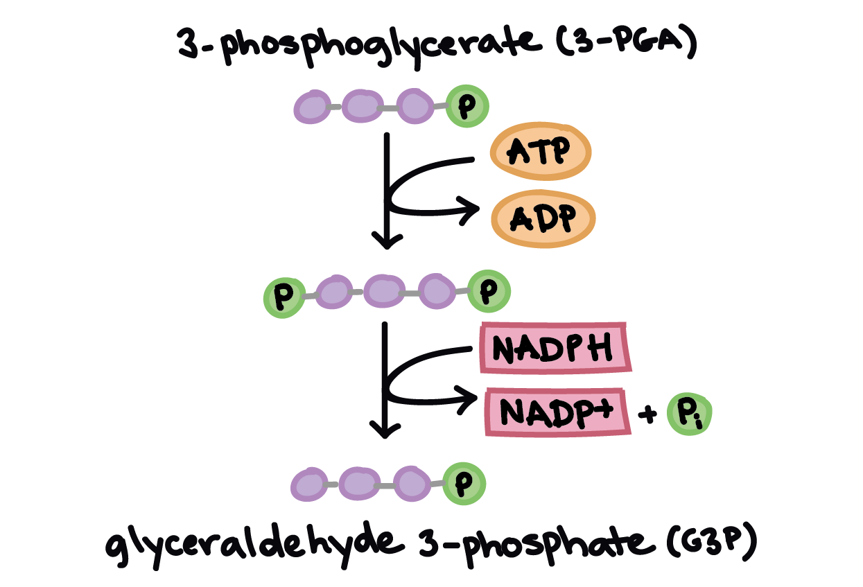
This compound is reduced to TP, or triose phosphate; as they are reduced (addition of H+), NADPH is oxidised to complete the redox reaction. Energy for this comes from ATP
Now we have 6 Triose phosphate; 1 goes to being 3 Carbon molecules of glucose molecule
Other 5 used in regeneration of RuBP
RuBP is a 5C compound
We have 5x3 compound
therefore we can make 3 RuBP;
energy from regeneration comes from ATP; complete the cycle
2 full cycles to make one glucose molecule
Chloroplast Structure and Function
adapted to its function in photosynthesis
Feature
double membrane (chloroplast envelope)
extensive internal membrane system (thylakoid)
small fluid spaces inside thylakoid
colourless fluid in chloroplast: stroma (many enzymes)
stacks of thylakoids: grana
strcuture/ function relationship
Structure
Function
chloroplasts absorb light
pigment molecules; arranged in photosystems in thylakoid membrane; large area of thylakoid ensure large surface area of absorption; stacks (Grana) allow more light to be absorbed
ATP by phosphorylation
proton gradient required between inside and outside of thylakoid; volume of fluid in it is small, so gradient can be built up quickly;
many chemical reaction during Calvin’s cycle
stroma has high concentration of required enzymes; ATP and reduced NADP, needed for the Calvin cycle, are easily available because the thylakoids, distributed through the stroma
Sources
Biology - Course Companion - Andrew Allott and David Mindorff - Oxford 2014
Chromatin looks like a string of beads because of lengths of DNA wound around histone proteins and separated by lengths of DNA.
G1: Cell enlarges.
S: Chromosomes replicate: there are two chromatids on each chromosome now; they are genetically identical.
G2: centrioles replicate.
Meiosis I
Prophase I
Homologous pairs pair up.
So there are four chromatids attached together.
This is called synapsis and the two pairs together make four chromatids.
The structure formed is called a bivalent or tetrad.
In eukaryotic cells, usually a synaptonemal complex is formed (protein structure that forms between homologous chromosomes (two pairs of sister chromatids).
Crossing over of homologous pairs occurs.
A junction is created where one chromatid in each homologous chromosome breaks and rejoins with a non-sister chromatid – occurs at random positions and can be several.
Chiasma: the point of contact between these two chromatids of homologous chromosomes. There can be many such points – chiasmata.
Importance of crossing over: increased stability of bivalents at chiasmata AND it creates new combinations of genes, also decoupled linked genes, leading to independent assortment.
Occurs at the same position on the two chromatids involved.
Chromosomes condense (shorten by supercoiling)
Nucleolus dissipates.
Centrioles migrate to the poles of the cell.
Spindle microtubules are growing from the poles of the cell.
Metaphase I
Spindle microtubules from the centrioles attach to the centromeres of the chromosomes.
The pole to which each chromosome is attached depends on which way the bivalent is facing – called orientation and it is random.
Bivalents align at the equator of the cell.
Nuclear envelope disintegrates.
Anaphase I
Microtubules shorten and pull bivalents apart and one chromosome of each pair moves to each pole. Important note: centromeres are not divided. This process is called disjunction.
Telophase I
Nuclear envelopes reforms.
Chromosomes uncoil.
Cytokinesis occurs and in the interphase between meiosis I and meiosis II and centrioles replicate but nothing else replicates.
Meiosis II
Prophase II
Chromosomes condense again.
Centrioles migrate to poles of the daughter cells as the spindle fibre network reforms.
Metaphase II
Nuclear envelope disintegrates.
Spindle microtubules attach themselves to the centromeres of the chromosomes.
Chromosomes align in the equator of the cell.
Anaphase II
Nuclear envelope disintegrates and chromatids are moved to opposite poles.
Haploid cells are created
Telophase II
Chromatids reach opposite poles.
Nuclear envelope reforms.
Cytokinesis occurs.
Meiosis and Genetic Variation
Mendel’s First Law: the Law of Segregation states that two alleles of every gene that occurs during meiosis separate (i e there is a 50% chance a particular allele is in a sex cell). Linked genes are more unlikely to separate because their loci are near each other.
Random orientation: bivalents face poles of the cell randomly and the orientation of one bivalent doesn’t affect that of another. It is the process that generates genetic variation among genes that are on different chromosome types.
Mendel’s Second Law: the Law of Independent Assortment states that the alleles of two genes will pass into gametes without influencing each other.
Crossing over: exchange of genetic materials forms new combinations (recombination).
Inability of a species to breed with a related species due to some factors (see below).
Only promotes selection in sexually reproducing organisms
Temporal Isolation
Mating seasons/times do not coincide
For example - different pollination times for flowers
Ecological Isolation
Organisms in the same area, but different habitats/conditions.
For example - Plant A survives in alkaline soil vs. Plant B in acidic
Behavioural Isolation
Organisms that mate based off of courting behaviour/pheromones will only mate with those who perform the best mating behaviour (eg dancing, fighting, etc.).
So it can prevent two organisms from mating.
Rate of Speciation
Phyletic Gradualism
Evolution occurring at a constant pace. Gradual Change.
Due to accumulation of mutations.
Punctuated Equilibrium
Long periods of stability followed by sudden changes
Fossil record supports this
Rapid evolution due to major environmental changes like a meteor
Polyploidy
Non-disjunction can occur during meiosis in humans.
Individual can end up with an extra chromosome or missing chromosomes.
Total non-disjunction: One of the two cells produced during Meiosis I gets all of the chromosomes.
Tetraploid offspring cannot mate with diploid organisms, so speciation has occurred
More common in plants
increased size, resistance to disease and overall vigour in plants.
Sources
Biology Class notes
Biology - Course Companion - Andrew Allott and David Mindorff - Oxford 2014